tandfonline | In portraiture,
subjects are mostly depicted with a greater portion of the left side of
their face (left hemiface) facing the viewer. This bias may be induced
by the right hemisphere's dominance for emotional expression and agency.
Since negative emotions are particularly portrayed by the left
hemiface, and since asymmetrical hemispheric activation may induce
alterations of spatial attention and action-intention, we posited that
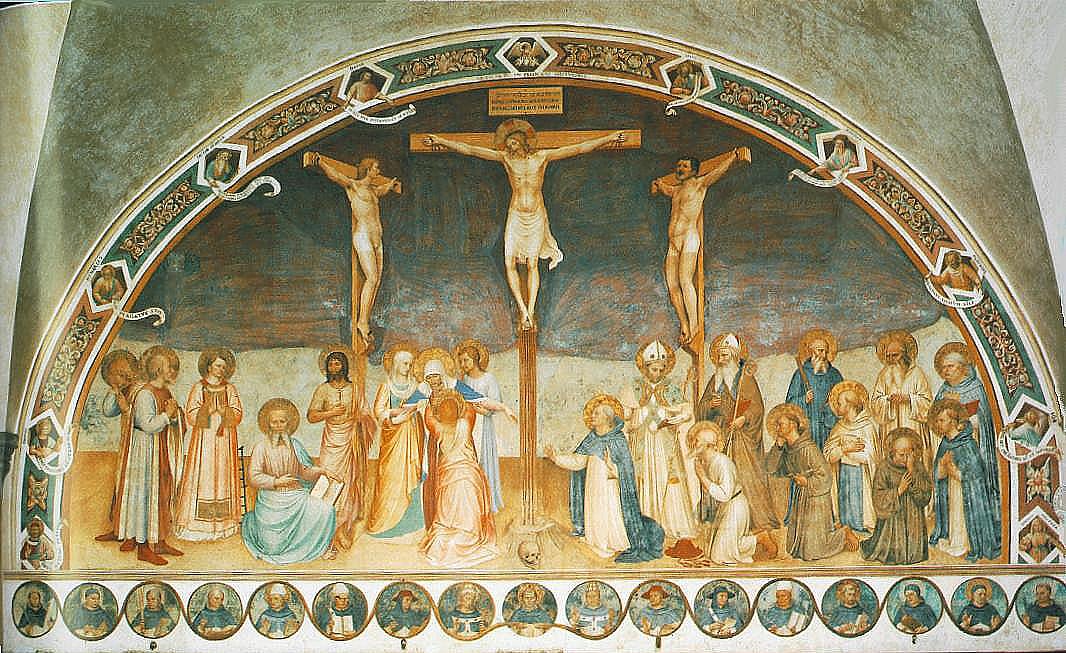
paintings of the painful and cruel crucifixion of Jesus would be more
likely to show his left hemiface than observed in portraits of other
people. By analyzing depictions of Jesus's crucifixion from book and art
gallery sources, we determined a significantly greater percent of these
crucifixion pictures showed the left hemiface of Jesus facing the
viewer than found in other portraits. In addition to the facial
expression and hemispatial attention-intention hypotheses, there are
other biblical explanations that may account for this strong bias, and
these alternatives will have to be explored in future research.
In
portraits, most subjects are depicted with their head rotated
rightward, with more of the left than right side of the subject's face
being shown. For example, in the largest study of facial portraiture,
McManus and Humphrey (
1973)
studied 1474 portraits and found a 60% bias to portray a greater
portion of the subjects’ left than right hemiface. Nicholls, Clode,
Wood, and Wood (
1999) found the same left hemiface bias even when accounting for the handedness of the painter.
Multiple
theories have been proposed in an attempt to explain the genesis of
this left hemiface bias in portraits. One hypothesis is that the right
hemisphere is dominant for mediating facial emotional expressions. In an
initial study, Buck and Duffy (
1980)
reported that patients with right hemisphere damage were less capable
of facially expressing emotions than those with left hemisphere damage
when viewing slides of familiar people, unpleasant scenes, and unusual
pictures. These right-left hemispheric differences in facial
expressiveness have been replicated in studies involving the spontaneous
and voluntary expression of emotions in stroke patients with focal
lesions (Borod, Kent, Koff, Martin, & Alpert,
1988; Borod, Koff, Lorch, & Nicholas,
1985; Borod, Koff, Perlman Lorch, & Nicholas,
1986; Richardson, Bowers, Bauer, Heilman, & Leonard,
2000).
Hemispheric
asymmetries are even reported in more “naturalistic” settings outside
the laboratory. For example, Blonder, Burns, Bowers, Moore, and Heilman (
1993)
videotaped interviews with patients and spouses in their homes and
found that patients with right hemisphere damage were rated as less
facially expressive than left hemisphere-damaged patients and normal
control patients. These lesion studies suggest that the right hemisphere
has a dominant role in mediating emotional facial expressions. Whereas
corticobulbar fibers that innervate the forehead are bilateral, the
contralateral hemisphere primarily controls the lower face. Thus, these
lesion studies suggest that the left hemiface below the forehead, which
is innervated by the right hemisphere, may be more emotionally
expressive.
This right hemisphere-left
hemiface dominance postulate has been further supported by studies of
normal subjects portraying emotional facial expressions. For example,
Borod et al. (
1988)
asked subjects to portray emotions, either by verbal command or visual
imitation. The judges who rated these facial expressions ranked the left
face as expressing stronger emotions. Sackeim and Gur (
1978)
showed normal subjects photographs of normal people facially expressing
their emotions and asked participants to rate the intensity of the
emotion being expressed. However, before showing these pictures of
people making emotional faces, Sackeim and Gur altered the photographs.
They either paired the left hemiface with a mirror image of this
photograph's left hemiface to form a full face made up of two left
hemifaces or formed full faces from right hemifaces. Normal participants
found that the composite photographs of the left hemiface were more
emotionally expressive than the right hemiface. Triggs, Ghacibeh,
Springer, and Bowers (
2005)
administered transcranial magnetic stimulation (TMS) to the motor
cortex of 50 subjects during contraction of bilateral orbicularis oris
muscles and analyzed motor evoked potentials (MEPs). They found that the
MEPs elicited in the left lower face were larger than the right face,
and thus the left face might appear to be more emotionally expressive
because it is more richly innervated.
Another
reason portraits often have the subjects rotated to the right may be
related to the organization of the viewer's brain. Both lesion studies
(e.g., Adolphs, Damasio, Tranel, & Damasio,
1996; Bowers, Bauer, Coslett, & Heilman,
1985; DeKosky, Heilman, Bowers, & Valenstein,
1980) and physiological and functional imaging studies (e.g., Davidson & Fox,
1982; Puce, Allison, Asgari, Gore, & McCarthy,
1996; Sergent, Ohta, & Macdonald,
1992)
have revealed that the right hemisphere is dominant for the recognition
of emotional facial expressions and the recognition of previously
viewed faces (Hilliard,
1973; Jones,
1979).
In addition, studies of facial recognition and the recognition of
facial emotional expressions have demonstrated that facial pictures
shown in the left visual field and left hemispace are better recognized
than those viewed on the right (Conesa, Brunold-Conesa, & Miron,
1995).
Since the right hemisphere is dominant for facial recognition and the
perception of facial emotions when viewing faces, the normal viewer of
portraits may attend more to the left than right visual hemispace and
hemifield. When the head of a portrait is turned to the right and the
observer focuses on the middle of the face (midsagittal plane), more of
the subject's face would fall in the viewer's left visual hemispace and
thus be more likely to project to the right hemisphere.
Agency
is another concept that may influence the direction of facial deviation
in portraiture. Chatterjee, Maher, Gonzalez Rothi, and Heilman (
1995)
demonstrated that when right-handed individuals view a scene with more
than one figure, they are more likely to see the left figure as being
the active agent and the right figure as being the recipient of action
or the patient. From this perspective the artist is the agent, and
perhaps he or she is more likely to paint the left hemiface of the
subject, which from the artist's perspective is more to the right, the
position of the patient. Support for this agency hypothesis comes from
studies in which individuals rated traits of left- versus right-profiled
patients, and found that those with the right cheek exposed were
considered more “active” (Chatterjee,
2002).
Taking
this background information into account and applying it to depictions
of the crucifixion of Jesus Christ highlights the various influences on
profile painting in portraiture. Specifically, we confirm the
predilection to display the left hemiface in portraiture and predict the
same in portraits of Jesus’ crucifixion.
The
strongest artistic portrayals of a patient being subject to cruel and
painful agents are images of the crucifixion of Jesus. The earliest
depiction of Christ on the cross dates back to around 420 AD. As
Christianity existed for several centuries before that, this seems to be
a late onset for this type of art. Because of the strong focus on
Christ's resurrection and the disgrace of his agony and death, art
historians postulate that there was a hesitation for early followers to
show Christ on the cross. The legalization of Christianity also may have
lifted the stigma. Based on the artwork still in existence from that
period, Jesus was often pictured alive during the crucifixion scene.
Several centuries later, from the end of the seventh century to the
beginning of the eighth century, Christ is more often shown dead on the
cross (Harries,
2005).
![]()
![]()
![]()
![]()
![]()
![]()
![]()
![]()
![]()
![]()
![]()
![]()
![]()


